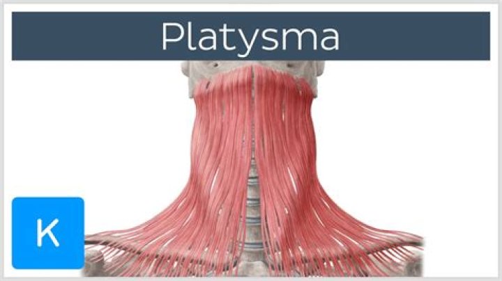
The platysma, innervated by the facial nerve, is a thin, sheet-like voluntary muscle. Origin: the muscle has a broad origin with fibers arising from the fascia of the upper thorax including the clavicle, acromial region, pectoralis major and deltoid muscles.
What artery supplies the platysma?
Results: The submental artery was the primary vessel to the platysma muscle. The superior thyroid artery, occipital artery, and posterior auricular artery were identified as secondary vessels. The external jugular vein provided the primary venous drainage, followed by the submental vein.
What movement does the platysma perform?
The actions of the platysma muscle include pulling down the mandible, which opens the mouth, and pulling the corners of the lips out to the side and down, which forms a frown. Additionally, the platysma muscle can form wrinkles in the neck as a person ages and their skin becomes less elastic and starts to sag.
What is the platysma muscle function?
The platysma is responsible for drawing the skin around the lower part of your mouth down or out, which creases the skin in your lower face, according to the Loyola University Medical Education Network.What is the antagonist of the platysma?
Platysma muscleAntagonistmasseter muscle, temporalis muscleIdentifiersLatinPlatysmaTA98A04.2.01.001
What supplies orbicularis oris?
Blood Supply and Lymphatics The main blood supply of this muscle is from the facial artery. Two of the branches of this artery, namely the superior labial branch and inferior labial branch, supply the muscle. It also receives supply from the maxillary artery via mental and infra-orbital branch.
What is the insertion of platysma?
OriginsSkin/fascia of infra- and supraclavicular regionsInsertionLower border of mandible, skin of buccal/cheek region, lower lip, modiolus, orbicularis oris muscleInnervationCervical branch of facial nerve (CN VII)Blood supplysubmental artery (facial artery), suprascapular artery (thyrocervical trunk)
How do you use platysma?
Pull the corners of your mouth downwards and outwards to create a contraction at the front of your neck. Relax, and repeat. This is the platysma muscle.What causes paralysis of the platysma?
Clinical relevance This is associated with the first signs of ageing of the neck, known as turkey neck. It occurs due to a decrease in muscle tone leading to thinning and shortening of the muscle. Platysma synkinesis can be a secondary complication of facial palsy.
What causes Platysmal banding?Platysmal bands are caused by aging and thickening of two edges of the neck muscles. There are many causes, including age, genetics, and muscle activity. The neck also has a much thinner layer of skin than the face.
Article first time published onWhere is the origin of platysma?
The platysma, innervated by the facial nerve, is a thin, sheet-like voluntary muscle. Origin: the muscle has a broad origin with fibers arising from the fascia of the upper thorax including the clavicle, acromial region, pectoralis major and deltoid muscles.
What is the Buccinator innervated by?
The buccinator is innervated by the buccal branches of facial nerve (CN VII).
What nerve innervates the orbicularis oculi?
The orbicularis oculi are innervated by the seventh cranial nerve, the facial nerve.
What is the Sternohyoid?
The sternohyoid is a strap like infrahyoid muscle that connects the hyoid bone with the clavicle and sternum. … The function of this muscle is to reestablish the breathing process by pulling the hyoid bone and larynx inferiorly after deglutition. This article will discuss the anatomy of the sternohyoid muscle.
What is the smallest muscle of the neck?
StapediusOriginWalls of pyramidal eminenceInsertionNeck of stapesArteryStapedial branch of posterior auricular arteryNerveFacial nerve (nerve to stapedius)
What is the most posterior neck muscle?
The trapezius muscles largely define the shape and outline of the neck, both from behind (here are the two trapezius muscles) and from in front. This is trapezius again. Trapezius is thought of mainly as a shoulder muscle. Its upper part raises the scapula.
Where is the Triangularis?
Muscles of the head, face, and neck (labeled as triangularis near chin). The depressor anguli oris muscle (triangularis muscle) is a facial muscle. It originates from the mandible and inserts into the angle of the mouth. It is associated with frowning, as it depresses the corner of the mouth.
What fascia forms a compartment for M Platysma?
The deep fascia of the neck lies deep to the superficial cervical fascia, a layer that is integral to the subcutaneous tissue and invests the platysma muscle.
Is the orbicularis oculi a sphincter?
Orbicularis oculi is considered the sphincter of the eyelids involved in facial expression, ocular protection and reflexes. Contraction of the orbital part draws the skin of the forehead and cheek towards the nose.
What is the origin of the orbicularis oris?
OriginMedial aspects of maxilla and mandible, perioral skin and muscles, modiolusInsertionSkin and mucous membrane of lipsActionCloses mouth, compresses and protrudes lipsInnervationBuccal branch of facial nerve (CN VII)
What Innervates the upper lip?
The infraorbital nerve, which is a terminal branch of the maxillary nerve, innervates the upper lip.
Where are the Frontalis?
Generally, the frontalis inserts at the eyebrow dermis and terminates laterally at the temporal ridge, but there is some variance and occasionally may terminate more medially as well. [10][11] While overall, it is a thin muscle with high vascularity, the bulk of it is located right above the brow.
How do you train a Platysma?
Use your tongue to help strengthen your platysma muscle. Sit up straight and open your mouth as far as you can, without discomfort. Stick your tongue out, then reach it down toward your chin. Hold for three to five seconds, then relax.
How do you remove Platysmal neck bands?
Plastic surgery is the primary treatment of this problem. A neck lift is a common procedure we perform to help get rid of the platysmal bands. This is a surgery that takes about two hours and requires an incision behind the ear and under the chin. In some people, an incision in front of the ear is also required.
What is medial pterygoid?
The medial pterygoid muscle, a major elevator of the jaw is a square-shaped masticatory muscle, located on the medial aspect of the lower jaw bilaterally. It is also known as internal pterygoid muscle. This muscle lies medial to the lateral pterygoid muscle.
What muscle elevates the mandible?
The function of the masseter muscle is to elevate the mandible and approximate the teeth—additionally, the intermediate and deep muscle fibers of the masseter function to retract the mandible.
What is the Buccinator mechanism?
Buccinator along with orbicularis oris and pharyngeal constrictor forms a functional unit (buccinator mechanism) which is essential for orofacial functions (swallowing, sucking, whistling, chewing, vowel pronunciation). … Also, the longitudinal fibers hold the bolus of food between the teeth during mastication.
What does the Buccinator attach to?
Buccinator muscleOriginfrom the alveolar processes of maxilla and mandible, buccinator crest and temporomandibular jointInsertionin the fibers of the orbicularis orisArterybuccal arteryNervebuccal branch of the facial nerve (VII cranial nerve)
What is the action of the Platysma quizlet?
What are the actions of the Platysma muscle? depresses the mandible and lower lip and tenses the skin of the anterior neck, shriek m. What nerve innervates the Platysma muscle? Where is the Sternocleidomastoid muscle?
What is levator Palpebrae Superioris innervated by?
The striated levator palpebrae superioris (LPS) muscle is innervated by the oculomotor nerve, and has a common origin with the superior rectus muscle. Anteriorly, it becomes the levator aponeurosis as it passes anterior to Whitnall ligament, and inserts into the anterior tarsal surface.
What is the levator Anguli Oris?
A muscle used in facial expression, primarily for smiling, the levator anguli oris elevates the angles of the mouth. The levator anguli oris originates roughly 1 cm inferior to the infraorbital foramen from the canine fossa of the maxilla and is located in the deepest layer of mimetic muscle.