Abstract. Extraoral radiography means that both the image detector and the X-ray machine are placed outside the patient’s mouth. The X-ray source and the image detector have to be aligned in order to generate the desired image quality.
What extraoral radiograph is used to best see the maxillary sinus?
Bregma–menton view. This projection is primarily used to demonstrate the walls of the maxillary sinus (especially in the posterior areas), the orbits, the zygomatic arches and the nasal septum.
What contains extraoral films during exposure?
Common type of Phosphor. Contains extraoral film during exposure. Shows the bony and soft tissue of the facial profile. … The part inside an extraoral cassette that converts x-ray energy into visible light, which in turn exposes screen film.
Which radiographs include types of intraoral radiographs?
- Bite-wing X-rays show details of the upper and lower teeth in one area of the mouth. …
- Periapical X-rays show the whole tooth — from the crown to beyond the end of the root to where the tooth is anchored in the jaw. …
- Occlusal X-rays are larger and show full tooth development and placement.
On which extraoral projections can the maxillary sinus be seen?
The maxillary sinus is sometimes referred to as the maxillary antrum and can be observed on both maxillary premolar and molar periapicals and partially on lateral-canine periapicals. Zygomatic bone – The zygomatic bone or cheek bone attaches to the right and left sides of the posterior maxilla.
What are the type of radiography?
There are three types of diagnostic radiographs taken in today’s dental offices — periapical (also known as intraoral or wall-mounted), panoramic, and cephalometric. Periapical radiographs are probably the most familiar, with images of a few teeth at a time captured on small film cards inserted in the mouth.
Which are the types of radiographic projection?
Basic radiographic projections include anteroposterior, where the X-ray beam enters the front of the body and exits through the back; posteroanterior, where it enters the back and exits the front; lateral projections, or side views; and oblique projections where the body is positioned at a 45-degree angle relative to …
What extraoral projection is used to evaluate fractures of the zygomatic arch?
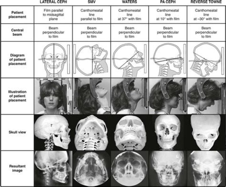
What projection is best for the examination of the maxillary sinusWaters ProjectionWhat projection is best for the examination of fractures of the zygomatic archSubmentovertex ProjectionWhat projection is best for the examination of fractures of the condylar neckReverse Towne ProjectionWhich type of film is more sensitive to light radiation intraoral or extraoral?
A screen film is more sensitive to fluorescent light than to direct exposure to X-rays. Nonscreen extraoral film is commonly used in extraoral radiography.
What is the lateral cephalometric projection used to evaluate?A lateral cephalometric radiograph (LCR) is a standardised, reproducible radiograph used primarily for orthodontic diagnosis and treatment planning. It is taken from a distance of 1.5m with the head at a right angle to the X-ray beam at a distance of 30cm, (although this has been found to vary slightly).
Article first time published onWhat is a panoramic radiograph used for?
Panoramic dental x-ray uses a very small dose of ionizing radiation to capture the entire mouth in one image. It is commonly performed by dentists and oral surgeons in everyday practice and may be used to plan treatment for dentures, braces, extractions and implants. This exam requires little to no special preparation.
What is intraoral and extraoral?
There are two main types of dental X-rays: intraoral (the X-ray film is inside the mouth) and extraoral (the X-ray film is outside the mouth). Intraoral X-rays are the most common type of X-ray.
What are the three types of intraoral imaging examinations?
Intraoral radiographic examination is the backbone of imaging for the general dental practitioner. It comprises of three categories: periapical, bitewing and occlusal projections.
Which type of intraoral projection is best for visualizing interproximal surfaces for decay?
In carious prone patients radiograph should be taken every 6 – 12 months; in non-caries prone patients every 18 – 24 months is adequate. The best radiographic view for visualizing both interproximal caries and periodontal bone height is bite-wing radiographs.
What are two types of extraoral film cassettes?
What size film is used for bitewing examinations? What are the two types of extraoral film cassettes? Cassettes are available in rigid and flexible styles.
How does extraoral film react differently from intraoral film?
How does extraoral film react differently from intraoral film? The light from the screen exposes the extraoral film; the intraoral film is exposed directly by radiation. … What is the primary difference between a film holder and a digital sensor holder? the size and shape of the holder.
What is a periapical image?
A periapical x-ray or “PA film” will show one or two teeth in their entirety in one single image, right from the crown of the tooth which is the part exposed in the mouth to the very tips of the tooth roots located in the jawbone, as well as the surrounding bone supporting this tooth.
Where are the maxillary sinuses?
A type of paranasal sinus (a hollow space in the bones around the nose). There are two large maxillary sinuses, one in each of the maxillary bones, which are in the cheek area next to the nose. The maxillary sinuses are lined with cells that make mucus to keep the nose from drying out.
What are maxillary landmarks?
Nasal fossae – The nasal fossae (plural; singular – fossa) are the nasal openings located above the maxillary anterior teeth. Nasal septum – The nasal septum is a bony vertical band-like midline structure that divides the nasal cavity into right and left chambers. …
Is the maxillary sinus radiopaque or radiolucent?
Maxillary sinus – a radiolucent area located above the apices of the maxillary premolars and molars. The floor of the maxillary sinus often appears as a thin wavy radiopaque line (bilateral).
Which are types of radiographic projection quizlet?
- Posteroanterior (PA) projection. The CR (central ray) enters the posterior surface and exits at the anterior surface.
- Anteroposterior (AP) projection. …
- AP oblique projection. …
- PA oblique projection. …
- Axial projection. …
- Inferosuperior axial projection. …
- Superoinferior axial projection. …
- Tangential projection.
What is a projection image?
An image projection occurs whenever a flat image is mapped onto a curved surface, or vice versa, and is particularly common in panoramic photography. A projection is performed when a cartographer maps a spherical globe of the earth onto a flat piece of paper, for example.
What are the two types of radiography?
Radiology may be divided into two different areas, diagnostic radiology and interventional radiology.
What does Projection mean in radiology?
Projection refers to the way the x-ray beam, like an arrow, passes through the body when the person is in that position. Remember, that arrow can pass through and project front to back, back to front, side to side, and so forth.
What is positioning in radiology?
Position denotes the placement of the patient’s body, specifically the portion of the patient’s anatomy that is in contact with the Bucky. For example, C indicates a lateral projection in a right lateral position, and D indicates a lateral projection in a left lateral position.
What is involved in radiography?
Undergraduates. Radiographers are healthcare professionals that use radiation for diagnosis and/or treatment of diseases. … They are also involved in interventional procedures, radiation Therapy, care of patients, management of radiography practice, research and Continued Professional Development (CPD).
How latent image is formed?
A latent image is an invisible image produced by the exposure to light of a photosensitive material such as photographic film. When photographic film is developed, the area that was exposed darkens and forms a visible image. … If intense exposure continues, such photolytic silver clusters grow to visible sizes.
Which of the following landmarks is useful when mounting films in the maxillary posterior area?
Which of the following landmarks is useful when mounting films in the maxillary posterior area? Floor of the maxillary sinus.
What is labial mounting?
Technique: Current convention is that all dental radiographs are mounted/interpreted with “labial mounting”. This means that the film is viewed from the outside in. … If you are interpreting a standard radiograph, the key to properly identifying the imaged side is the embossed dot, which is on one corner of the film.
Which of the following film sizes is the most commonly used?
35mm film is easily the most popular choice. It was first made available in 1934. Most people are familiar with this format, it comes as a cartridge that fits into all 35mm film cameras. It is the easiest way to shoot film, it is the most portable and the most common.
Which of the following projection is best for examination of fractures of the mandibular body?
CT is the current diagnostic tool of choice for the radiographic evaluation and diagnosis of mandible fractures. A panoramic tomographic view (Panorex view) [6, 7, 4] shows the entire mandible in one plane.