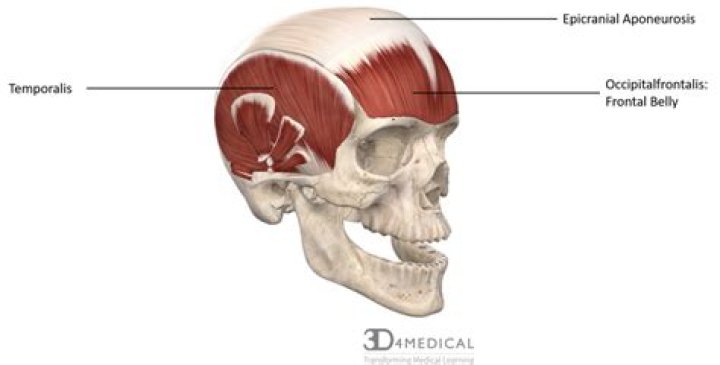
Epicranial Aponeurosis also referred to as the Galea Aponeurotica or the aponeurosis epicranialis is a broad tendon and is the intermediate section of the occipitofrontalis muscle. It runs from the superior portion of the frontal bone and covers the parietal bones to the lambdoid suture.
What is the function of the epicranial aponeurosis?
The epicranial aponeurosis provides the insertion point for the occipitofrontalis muscle, a thin, broad muscle that covers the top of your skull. This muscle controls many of your facial expressions. Every time you raise your eyebrows, you can thank your occipitofrontalis muscle and your epicranial aponeurosis!
What does the Epicranial muscle do?
They insert on the mastoid process of the temporal bone. They can flex or extend the head, or can rotate the towards the shoulders. The epicranius muscle is also very broad and covers most of the top of the head. The epicranius muscle includes a middle section which is all aponeurosis.
What is the origin of the epicranial aponeurosis?
Structure. In humans, the epicranial aponeurosis originates from the external occipital protuberance and highest nuchal lines of the occipital bone. It merges with the occipitofrontalis muscle.What is the function of an aponeurosis?
1. A: aponeuroses are extensions of external tendons on the surface of pennate muscles that function as insertion sites for muscle fascicles and may play a role in modulating fascicle rotation and dynamic gearing during muscle contractions.
What is the function of the orbicularis oculi?
Structure and Function The orbicularis oculi muscle closes the eyelids and assists in pumping the tears from the eye into the nasolacrimal duct system. The orbital section of the orbicularis oculi is more involved in the voluntary closure of the eyelid, such as with winking and forced squeezing.
What does the word aponeurosis mean in medical terms?
aponeurosis, a flat sheet or ribbon of tendonlike material that anchors a muscle or connects it with the part that the muscle moves. … Aponeuroses are structurally similar to tendons and ligaments.
What is the action of Frontalis?
FrontalisActionsRaises eyebrows and wrinkles foreheadIdentifiersLatinVenter frontalis musculi occipitofrontalisTA98A04.1.03.004Where is the Epicranial Aponeurosis located?
Epicranial Aponeurosis also referred to as the Galea Aponeurotica or the aponeurosis epicranialis is a broad tendon and is the intermediate section of the occipitofrontalis muscle. It runs from the superior portion of the frontal bone and covers the parietal bones to the lambdoid suture.
What is occipital belly?The occipitalis muscle (occipital belly) is a muscle which covers parts of the skull. Some sources consider the occipital muscle to be a distinct muscle. … The occipitalis muscle is innervated by the facial nerve and its function is to move the scalp back. The muscles receives blood from the occipital artery.
Article first time published onWhat is Buccinator muscle?
The buccinator muscle plays an active role along with orbicularis oris and superior constrictor muscle during swallowing, mastication, blowing, and sucking. It aids in mastication and blowing by compressing the cheek inwards.
What is the origin of the orbicularis oculi muscle?
OriginNasal part of frontal bone, frontal process of maxilla, medial palpebral ligament, lacrimal boneInnervationTemporal and zygomatic branches of facial nerve (CN VII)Blood supplyMaxillary, superficial temporal, facial and ophthalmic arteries.
Where is the Triangularis?
Muscles of the head, face, and neck (labeled as triangularis near chin). The depressor anguli oris muscle (triangularis muscle) is a facial muscle. It originates from the mandible and inserts into the angle of the mouth. It is associated with frowning, as it depresses the corner of the mouth.
Why is aponeurosis different from tendon?
Key Difference The main difference is that Aponeurosis connects the muscles of the body to other muscles which necessitate help, while the tendons serve as a link between the muscles and the bones.
What is pulmonary aponeurosis?
FMA. 42435. Anatomical terminology. The palmar aponeurosis (palmar fascia) invests the muscles of the palm, and consists of central, lateral, and medial portions.
What's the difference between a tendon and aponeurosis?
Aponeurosis is an extremely delicate, thin sheath-like structure, which attaches muscles to the bones whereas tendons are tough, rounded cord-like structures which are extensions of the muscle. Normally, tendons allow the attachment of the muscle from its originating bone to the bone on which it ends.
Is the linea alba an aponeurosis?
The linea alba is an aponeurosis of the ventral muscles and plays a bigger role for the rectus sheath.
What is Bicipital Aponeurosis?
The bicipital aponeurosis (also known as lacertus fibrosus) is a broad aponeurosis of the biceps brachii, which is located in the cubital fossa of the elbow. It separates superficial from deep structures in much of the fossa.
Which is an example of an aponeurosis quizlet?
An aponeurosis is a broad sheet of dense connective tissue that connects a muscle to another muscle or to bone. An example of an aponeurosis is the galea aponeurotica, the origin of the frontalis. Skeletal muscles are covered by three continuous layers of connective tissue.
What kind of muscle is the orbicularis oculi in terms of action?
Orbicularis oculi muscleActionscloses eyelidsAntagonistlevator palpebrae superiorisIdentifiersLatinmusculus orbicularis oculi also musculus orbicularis palpebrarum
What happens if orbicularis oculi is damaged?
If the orbicularis oculi muscles were damaged, you would not be able to blink or wink. Also, humans often express emotion though their eyes and this would be impaired.
Which part of the orbicularis oculi is known as Horner's muscle?
Horner’s muscle (the palpebral part of the orbicularis oculi muscle) has a fan-shaped origin in the lacrimal bone. Its muscle fibers are oriented from 160 to 210 degrees relative to the ear-eye plane and converge towards the medial palpebral commissure.
What type of tissue is the epicranial aponeurosis?
The galea aponeurotica (also called the galeal or epicranial aponeurosis or the aponeurosis epicranialis) is a tough fibrous sheet of connective tissue that extends over the cranium, forming the middle (third) layer of the scalp.
What is an aponeurosis quizlet?
An aponeurosis is a broad fibrous sheet of connective tissue that connects muscles to adjacent muscles. … Muscle fibers are grouped together in fascicles that are surrounded by a layer of connective tissue called the endomysium.
What type of muscle is Frontalis?
The frontalis muscle is a thin, wide, four-sided muscle located at the top front of the skull (in the area of the forehead). Specifically, this muscle originates from the galea aponeurotica and extends down the forehead and inserts or attaches to the skin around the eyebrows and top of the nose.
What is the Frontalis origin?
The origin of the frontalis is at the hairline, known as the epicranium of the aponeurosis. Its insertion is at the level of the eyebrows, where it is intertwined with the fibers of the procerus, corrugator, depressor supercilii, and orbicularis oculi muscles.
How many muscles are in the Frontalis?
The frontalis muscles are two large fanlike muscles that extend from the eyebrow region to the top of the forehead.
How do you relax the occipital muscles?
Apply gentle pressure from your fingertips at the base of your skull. This massage can help calm tight muscles and release tension. You can also place a rolled towel under your head and neck as you lie down on your back. The pressure from the towel can provide a gentle massage.
Can you cure occipital neuralgia?
Treatment of occipital neuralgia aims to alleviate the pain; however, it is not a cure. Interventions can be surgical or non-surgical.
What is Zygomaticus major?
Of all the muscles in the face, the zygomaticus major is perhaps the most noticeable. Sitting between the corners of our lips and the upper part of our cheeks, it controls the way in which we smile. The muscle sits atop the zygomatic bone, otherwise known as the cheekbone.
What is the Buccinator origin?
ORIGIN. External alveolar margins of maxilla and mandible by molar teeth, to maxillary tubercle and pterygoid hamulus and posterior mylohyoid line respectively, then via pterygomandibular raphe between bones. INSERTION.