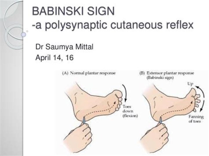
In adults or children over 2 years old, a positive Babinski sign happens when the big toe bends up and back to the top of the foot and the other toes fan out. This can mean that you may have an underlying nervous system or brain condition that’s causing your reflexes to react abnormally.
Is a positive Babinski reflex bad?
The reflex may be present in infants without any underlying conditions. After the age of 2 years, though, the Babinski reflex should be absent. A positive result in adults or children over the age of 2 years may be a sign of an underlying issue in the central nervous system.
What is normal Babinski reflex?
Babinski reflex When the sole of the foot is firmly stroked, the big toe bends back toward the top of the foot and the other toes fan out. This is a normal reflex up to about 2 years of age.
When is a positive Babinski normal?
This reflex is normal in children up to 2 years old. It disappears as the child gets older. It may disappear as early as 12 months.Why is Babinski positive in infants?
The Babinski response is a primitive reflex which occurs because the corticospinal pathways (bundles of nerve fibers) running from the brain and down the spinal cord are not fully myelinated (sheathed) in newborns and infants.
Why is Babinski positive in UMN lesions?
In Babinski’s sign, there is dorsiflexion of the big toe and abduction of the other toes. Physiologically, it is normally present in infants from birth to 12 months. The presence of the Babinski sign after 12 months is the sign of a non-specific upper motor neuron lesion. Increased deep tendon reflex (DTR)
Is Babinski reflex present at birth?
However, other reflexes are unique to infants, and they typically grow out of these reflexes within a few months of birth. These reflexes include: asymmetrical tonic neck reflex. Babinski reflex.
What are the five infant reflexes?
- Rooting reflex. This reflex starts when the corner of the baby’s mouth is stroked or touched. …
- Suck reflex. Rooting helps the baby get ready to suck. …
- Moro reflex. The Moro reflex is often called a startle reflex. …
- Tonic neck reflex. …
- Grasp reflex. …
- Stepping reflex.
What is the difference between plantar and Babinski reflex?
The differences between these two reflexes are in the receptive fields and the fact that the great toe is flexed in one and extended in the other. … The abnormal plantar reflex, or Babinski reflex, is the elicitation of toe extension from the “wrong” receptive field, that is, the sole of the foot.
What are the 7 reflexes of a newborn?- Moro Reflex. Babies usually exhibit a full Moro reflex which includes the arms, head and legs in their first 12 weeks after birth. …
- Rooting Reflex. …
- Sucking Reflex. …
- Tonic Neck Reflex. …
- Grasp Reflex. …
- Babinski Reflex. …
- Stepping Reflex.
Why do upper motor neuron lesions cause spasticity?
How does UMN lesion cause spasticity and associated phenomena? The major problem is a loss of control of the spinal reflexes. Spinal reflex activity is normally tightly regulated and if inhibitory control is lost, the balance is tipped in favor of excitation, resulting in hyperexcitability of the spinal reflexes.
What causes upper motor neuron signs?
Presentation. The upper motor neuron syndrome signs are seen in conditions where motor areas in the brain and/or spinal cord are damaged or fail to develop normally. These include spinal cord injury, cerebral palsy, multiple sclerosis and acquired brain injury including stroke.
Why do upper motor neuron lesions cause Hyperreflexia?
Hyperreflexia. Because of the loss of inhibitory modulation from descending pathways, the myotatic (stretch) reflex is exaggerated in upper motor neuron disorders. The stretch reflex is a major clinical diagnostic test of whether a motor disorder is caused by damage to upper or lower motor neurons.
What is Downgoing Babinski?
This abnormal finding suggests a lesion of the corticospinal tract (upper motor neurons) in the brain, brainstem or spinal cord. The normal response to stroking the sole of the foot is flexion of the toes (downgoing toes).
What is the normal plantar response?
The normal plantar reflex consists of flexion of the great toe or no response. … A positive Chaddock sign refers to dorsiflexion of the great toe after stroking from the lateral ankle to the lateral dorsal foot. A positive Stransky sign refers to an upgoing great toe after flipping the little toe outward.
What muscle's contract during a normal plantar reflex which are relaxed?
Gastrocnemius: This muscle makes up half of your calf muscle. It runs down the back of your lower leg, from behind your knee to the Achilles tendon in your heel. It’s one of the main muscles involved in plantar flexion. Soleus: The soleus muscle also plays a major role in plantar flexion.
What causes knee jerk?
The normal knee-jerk or, “patellar jerk,” reflex is elicited when the knee is tapped below the knee cap (patella). Sensors that detect stretching of the tendon of this area send electrical impulses back to the spinal cord.
How can I test my reflexes without a hammer?
When checking knee reflexes, press down on the dorsum of the foot while tapping the patellar tendon. This maneuver overcomes inhibition of the reflex, so that a brisk tap with the side of the index finger elicits a good response.
Where is caput Succedaneum located?
A caput succedaneum refers to a predominantly serous or occasionally a serous-sanguineous fluid collection within the scalp located in the compartment between skin and galea or epicranial aponeurosis. A caput succedaneum typically results from high pressure exerted on the infant’s head during labor.
Who is the Babinski reflex named after?
The Babinski reflex was described by the neurologist Joseph Babinski in 1899. Since that time, it has been incorporated into the standard neurological examination.
What is the meaning of Babinski?
Definition of Babinski reflex : a reflex movement in which when the sole is tickled the big toe turns upward instead of downward and which is normal in infancy but indicates damage to the central nervous system (as in the pyramidal tracts) later in life.
What is rooting in a baby?
The rooting reflex happens when the corner of a baby’s mouth touches the skin or nipple. You can also trigger the reflex by stroking or gently touching the corner of a baby’s mouth. A baby will then reflexively turn their head to follow and “root” in that direction.
What is plantar grasp?
The plantar grasp reflex (Babinski reflex) is similar to the grasp reflex of the hand. If you place your thumb below the toe bed of an infant’s foot and apply pressure, the toes will curl around your thumb, grasping it (flexion and adduction). This reflex is not present in many newborns.
What is the purpose of Moro reflex?
Function. The Moro reflex may be a survival instinct to help the infant cling to its mother. If the infant lost its balance, the reflex caused the infant to embrace its mother and regain its hold on the mother’s body.
Is sneezing a newborn reflex?
Sneezing in infants is a reflex just like it is with adults. The reflex occurs when the nasal passages are irritated. Unlike a lot of other reflexes like the startle reflex or the Moro reflex, the sneezing reflex is one that sticks around as the baby grows and into adulthood.
Is Moro reflex normal?
These reflexes are a normal part of a baby’s development. They help your baby function in the world. The Moro reflex is another normal baby reflex.
Why do babies hold fingers?
The grasp reflex is an involuntary movement that your baby starts making in utero and continues doing until around 6 months of age. It’s a crowd-pleaser of a reflex: This is the reflex at play when your newborn wraps their adorable little fingers around one of yours.
What is Babinski and clonus?
Sustained clonus does not stop as long as dorsiflexion pressure is applied to the foot. Babinski reflex: for loss of brain control over lower extremities; scraping the soles causes toes to pull up.
How can you tell the difference between upper and lower motor neuron lesions?
An upper motor neuron lesion is a lesion of the neural pathway above the anterior horn of the spinal cord or motor nuclei of the cranial nerves. A Lower motor neuron lesion is a lesion which affects nerve fibers traveling from the anterior horn of the spinal cord to the associated muscle(s).
How can you tell the difference between UMN and LMN lesions?
Although both upper and motor neuron lesions result in muscle weakness, they are clinically distinct due to various other manifestations. Unlike UMNs, LMN lesions present with muscle atrophy, fasciculations (muscle twitching), decreased reflexes, decreased tone, negative Babinsky sign, and flaccid paralysis.
What is Brown Séquard syndrome?
Brown-Séquard syndrome is a rare spinal disorder that results from an injury to one side of the spinal cord in which the spinal cord is damaged but is not severed completely. It is usually caused by an injury to the spine in the region of the neck or back.