The inner surface of the gall bladder is covered by the mucosa. The sufrace is made up of a simple columnar epithelium. The epithelial cells have microvilli, and look like absorptive cells in the intestine. Underneath the epithelium is the lamina propria.
Which of the following is lined by a mucosa?
Mucous membranes line many tracts and structures of the body, including the mouth, nose, eyelids, trachea (windpipe) and lungs, stomach and intestines, and the ureters, urethra, and urinary bladder.
What layers are not found in the gallbladder?
Underneath the epithelium is an underlying lamina propria, a muscular layer, an outer perimuscular layer and serosa. Unlike elsewhere in the intestinal tract, the gallbladder does not have a muscularis mucosae, and the muscular fibres are not arranged in distinct layers.
Does gallbladder have Muscularis mucosa?
Based on our findings, we conclude that, in the gallbladder wall, the muscle layer is muscularis propria and there is no muscularis mucosae present.What is the gallbladder fossa?
Description. The fossa for the gall-bladder (fossa vesicæ felleæ) is a shallow, oblong fossa, placed on the under surface of the right lobe, parallel with the left sagittal fossa. It extends from the anterior free margin of the liver, which is notched by it, to the right extremity of the porta.
What is stomach mucosa?
The gastric mucosa is the mucous membrane layer of the stomach, which contains the glands and the gastric pits. In humans, it is about 1 mm thick, and its surface is smooth, soft, and velvety. It consists of simple columnar epithelium, lamina propria, and the muscularis mucosae.
What is a Cholangiocyte?
Cholangiocytes are a heterogeneous, highly dynamic population of epithelial cells that line a three-dimensional network of bile ducts known as the biliary tree. Their major physiologic function lies in modification of hepatic canalicular (i.e., primary) bile as it is transported along the biliary tree.
What is mucosal tissue?
n. A membrane lining all body passages that communicate with the air, such as the respiratory and alimentary tracts, and having cells and associated glands that secrete mucus. Also called mucosa.What type of cells produce the mucus for the mucous membranes?
Goblet cells produce the mucus for the mucous membranes.
Does gall bladder have villi?The gall bladder has a wrinkled mucosa; sections across the wrinkles superficially resemble villi.
Article first time published onWhat Innervates the gallbladder?
The gallbladder receives parasympathetic nerve supply from the right vagus through its hepatic branch; sympathetic supply comes from T 7-9 through the celiac plexus.
Where is muscularis mucosa found?
The lamina muscularis mucosae (or muscularis mucosae) is a thin layer (lamina) of muscle of the gastrointestinal tract, located outside the lamina propria, and separating it from the submucosa.
Why is submucosa absent in gallbladder?
SUBMUCOSA OF GALL BLADDER A MISCONCEPTION Muscularis mucosa is totally absent in the wall of the gall bladder. The lamina propria containing loose connective tissue rest upon the muscularis externa and hence there is no muscular layer separating the mucosa from the muscularis externa.
What is Phrygian cap of gallbladder?
A Phrygian cap is a congenital anomaly of the gallbladder with an incidence of 4%. It can simulate a mass in the liver during hepatobiliary imaging and is sometimes mistaken for pathology. A Phrygian cap, however, has no pathological significance and normally causes no symptoms.
Which type of epithelium is present in bile duct?
The extrahepatic bile ducts are lined by high columnar epithelial cells and together with the large intrahepatic bile ducts contain the PBGs [15].
What is Calots triangle?
The triangle of Calot is an important landmark whose boundaries include the common hepatic duct medially, the cystic duct laterally, and the inferior edge of the liver superiorly. … This triangular space is dissected to allow the surgeon to identify, divide, and ligate the cystic duct and artery.
What liver segment is the gallbladder in?
The gallbladder straddles the undersurfaces of liver segments IVB and V. There is an H-shaped fissure on the inferior surface of the liver. The right vertical arm of the H is formed by the gallbladder anteriorly and the inferior vena cava (IVC) posteriorly; it is incomplete, with the caudate process between the two.
Is the gallbladder fossa part of the gallbladder?
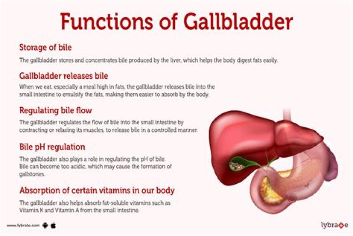
The gallbladder, which stores bile produced by the liver, normally is located in the gallbladder fossa, a depression on the visceral surface of the liver located between the right and quadrate anatomical liver lobes [1].
What is a stellate cell?
The stellate cell, previously known as the Ito cell, fat-storing cell, perisinusoidal cell or lipocyte, is a major storage site for vitamin A. In liver injury, it becomes a transitional cell or myofibroblast-like cell capable of synthesising collagen types I, III and IV as well as laminin.
What is the hepatic triad?
por·tal tri·ad. (pōr’tăl trī’ad) Branches of the portal vein, hepatic artery, and the biliary ducts bound together in the perivascular fibrous capsule or portal tract as they ramify within the substance of the liver.
What is a liver sinusoid?
Sinusoids are low pressure vascular channels that receive blood from terminal branches of the hepatic artery and portal vein at the periphery of lobules and deliver it into central veins. Sinusoids are lined with endothelial cells and flanked by plates of hepatocytes.
What is fundic mucosa?
The large quantity of gastric fluid produced by the mammalian stomach is thought to be secreted mainly by fundic glands in the mucosa of the stomach body. These glands contain mucous cells, chief cells, and parietal cells that secrete mucus, pepsinogen, and hydrochloric acid, respectively.
What do the cells of the stomach mucosa produce?
Mucous cells: secrete an alkaline mucus that protects the epithelium against shear stress and acid. Parietal cells: secrete hydrochloric acid. Chief cells: secrete pepsin, a proteolytic enzyme.
What is released from gastric mucosa?
Gastric Secretions The stomach mucosa contains oxyntic or gastric glands and pyloric glands. Oxyntic glands secrete hydrochloric acid, pepsinogen, intrinsic factor and mucus; and pyloric glands secrete mucus and the hormone gastrin.
What is the mucosa made of?
The mucosa consists of epithelium, an underlying loose connective tissue layer called lamina propria, and a thin layer of smooth muscle called the muscularis mucosa. In certain regions, the mucosa develops folds that increase the surface area. Certain cells in the mucosa secrete mucus, digestive enzymes, and hormones.
Is mucous membrane epithelial or connective?
Mucous membranes are epithelial membranes that consist of epithelial tissue that is attached to an underlying loose connective tissue. These membranes, sometimes called mucosae, line the body cavities that open to the outside. The entire digestive tract is lined with mucous membranes.
Is mucosa an epithelium?
The mucosa is the inner layer of any epithelially-lined hollow organ (e.g., mouth, gut, uterus, trachea, bladder, etc.). The mucosa consists of the epithelium itself and also the supporting loose connective tissue, called lamina propria, immediately beneath the epithelium.
What cells are found in mucosa tissue?
The mucosa is the innermost layer, and functions in absorption and secretion. It is composed of epithelium cells and a thin connective tissue. The mucosa contains specialized goblet cells that secrete sticky mucus throughout the GI tract.
What is mucosal epithelial cell?
A mucous membrane or mucosa is a membrane that lines various cavities in the body and covers the surface of internal organs. It consists of one or more layers of epithelial cells overlying a layer of loose connective tissue.
What is an epithelial membrane?
The epithelial membrane is composed of epithelium attached to a layer of connective tissue, for example, your skin. The mucous membrane is also a composite of connective and epithelial tissues. … These membranes line cavities that do not open to the outside, and they cover the organs located within those cavities.
Where is the sphincter of Oddi?
The sphincter of Oddi refers to the smooth muscle that surrounds the end portion of the common bile duct and pancreatic duct. This muscle relaxes during a meal to allow bile and pancreatic juice to flow into the intestine.