There is no real cure or treatment for optic atrophy. Therefore, it’s important to have regular eye exams (especially if you have a family history of eye diseases), and to see your ophthalmologist immediately if you have any changes in your vision.
Can optic atrophy be cured?
There is no real cure or treatment for optic atrophy. Therefore, it’s important to have regular eye exams (especially if you have a family history of eye diseases), and to see your ophthalmologist immediately if you have any changes in your vision.
Can optic atrophy reversed?
Damage from optic nerve atrophy cannot be reversed. The underlying disease must be found and treated. Otherwise, vision loss will continue. Rarely, conditions that lead to optic atrophy may be treatable.
How common is optic atrophy?
Optic atrophy type 1 is estimated to affect 1 in 35,000 people worldwide. This condition is more common in Denmark, where it affects approximately 1 in 10,000 people.What diseases cause optic nerve atrophy?
- Brain tumor.
- Cranial arteritis (sometimes called temporal arteritis)
- Multiple sclerosis.
- Stroke.
Does optic atrophy show up on MRI?
[19] The diagnosis is based on opthalmoscopic findings. MRI reveals diffuse bilateral optic nerve atrophy.
Does optic atrophy get worse?
In general, people with optic atrophy type 1 have worsening vision loss over time. However, some people only have mild vision loss, and for some people the vision loss does not worsen with time.
Is optic atrophy painful?
The main symptom of optic atrophy is vision loss. Any other symptoms are attributable to the underlying process that caused the disc damage (such as pain with angle closure glaucoma).Is optic nerve atrophy progressive?
Optic nerve damage is usually permanent and in some cases progressive. By the time optic atrophy is detected, substantial optic nerve injury has already occurred.
What are signs of optic nerve damage?- Abnormal pupil size and nonreactivity to light.
- Bulging of the eyes.
- Complete or partial loss of vision.
- Diminished ability to see fine details.
- Diminished color vision or colors seem faded.
- Dimming or blurring of vision.
- Double vision.
- Eye redness.
What age group is affected by optic atrophy?
Optic atrophy type 1 (OPA1, or Kjer type optic atrophy) is characterized by bilateral and symmetric optic nerve pallor associated with insidious decrease in visual acuity (usually between ages 4 and 6 years), visual field defects, and color vision defects.
How do I stop my optic nerve from thinning?
Unfortunately, there is no effective treatment for optic atrophy. Once the nerve fibers in the optic nerve are lost they never heal or grow back. However, early diagnosis and treatment of the underlying causes of optic atrophy can help prevent further damage from the disease.
Is optic nerve atrophy degenerative?
Optic atrophy is the final common morphologic endpoint of disease process that causes degeneration of axons of the ganglion cells.
How is optic nerve damage treated?
- For people diagnose with glaucoma, treatment may involve use of eye drops, oral medications or getting eye surgeries like laser therapy or drainage tubes.
- For people suffering from Optic Nerve drusen, may benefit from medication that lowers intraocular pressure.
Does glaucoma show up on MRI?
Quantitative MRI parametric evaluation of GMD can detect glaucoma-associated anatomical atrophy of the visual cortex in BA 17, 18, and 19. Furthermore, GMD in BA 19 was significantly correlated to the damage level of the optic nerve, as well as the retina, in patients with OAG.
Where is the ocular nerve?
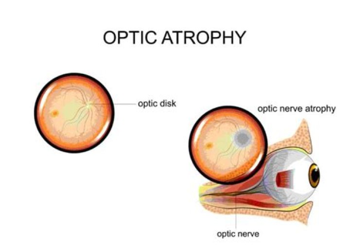
The optic nerve begins at the optic disk, a structure that is 1.5 mm (0.06 inch) in diameter and is located at the back of the eye. The optic disk forms from the convergence of ganglion cell output fibres (called axons) as they pass out of the eye.
What is Foster Kennedy syndrome?
Foster-Kennedy Syndrome is characterized by unilateral visual loss with a compressive optic atrophy in one eye and contralateral papilledema caused by increased intracranial pressure. The same ophthalmoscopic features however can be seen in the pseudo-Foster-Kennedy Syndrome.
What vitamin is good for the optic nerve?
Niacin. The main function of niacin (vitamin B3) in your body is to help convert food into energy. It can also act as an antioxidant (22). Recently, studies have suggested that niacin may play a role in the prevention of glaucoma, a condition in which the optic nerve of your eye becomes damaged (23).
Can stress cause optic neuritis?
In fact, continuous stress and elevated cortisol levels negatively impact the eye and brain due to autonomous nervous system (sympathetic) imbalance and vascular dysregulation; hence stress may also be one of the major causes of visual system diseases such as glaucoma and optic neuropathy.
How do doctors test the optic nerve?
Ophthalmoscopy. During this examination, your doctor shines a bright light into your eye and examines the structures at the back of your eye. This eye test evaluates the optic disk, where the optic nerve enters the retina in your eye.
What is dominant atrophy?
Autosomal dominant optic atrophy and cataract is an eye disorder that is characterized by impaired vision. Most affected individuals have decreased sharpness of vision (visual acuity) from birth, while others begin to experience vision problems in early childhood or later.
What are types of optic atrophy?
Isolated: These include dominant and recessive optic atrophy, Leber’s hereditary optic neuropathy, and Behr’s hereditary optic atrophy. Optic atrophy associated with systemic disease or neurological conditions.
Can optic nerve heal itself?
Damage to the optic nerve is irreversible because the cable of nerve fibers doesn’t have the capacity to regenerate, or heal itself, when damage occurs. This is why glaucoma is an incurable disease at this point, and why early detection is so important.