Papillary thyroid carcinoma.Papillary renal cell carcinoma.Ovarian papillary serous cystadenoma and cystadenocarcinoma.Endometrial adenocarcinomas (Papillary serous carcinoma ~3%-4%)Meningiomas, in the central nervous system.
What causes cervix calcification?
The most common causes of calcific endometritis are post-abortion (causing chronic inflammation from retained tissue after first and second trimester abortion) and chronic endometritis (due to genital TB, non-specific chronic endometritis and pyometra).
How do you know if you have a Psammoma body?
Psammoma bodies—from both non-cancerous and cancerous conditions—can be detected after a biopsy of the mass is taken and stained with haematoxylin and eosin, a principle tissue stain used in histology. Ultrasound may also be used to detect calcifications of thyroid nodules.
What is Psammomatous meningioma?
[ sə-mō′mə-təs ] n. A firm fibrous neoplasm of meninges of the brain and spinal cord characterized by psammona bodies. sand tumor.What causes Psammomatous calcification?
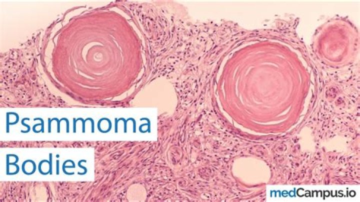
Psammoma bodies are characteristically associated with non-neoplastic serous proliferations and with serous neoplasms, which may be benign, borderline, or malignant. They are thought to arise secondary to necrosis, with subsequent dystrophic calcification of the tips of papillary structures.
What are the symptoms of calcification?
- Bone pain.
- Bone spurs (occasionally visible as lumps under your skin)
- Breast mass or lump.
- Eye irritation or decreased vision.
- Impaired growth.
- Increased bone fractures.
- Muscle weakness or cramping.
- New deformities such as leg bowing or spine curvature.
Are all cancers carcinomas?
Not all cancers are carcinoma. Other types of cancer that aren’t carcinomas invade the body in different ways. Those cancers begin in other types of tissue, such as: Bone.
What causes pelvic calcification?
Pelvic phleboliths may also be caused by an uncommon condition known as venous malformation, which results in abnormal development of veins. These veins stretch or enlarge over time. The blood circulates very slowly, resulting in blood clots that calcify over time to create phleboliths.What does it mean to have calcification in the uterus?
Calcification inside the uterine cavity is an uncommon finding that is usually related to secondary infertility. Although a few cases can be caused by osseous metaplasia, heteroplasia, or dystrophic calcification of the endometrium, the most common feature is a history of pregnancy loss or termination [1].
What cells do meningioma arise from?Meningiomas are the most common benign intracranial tumor. They originate from arachnoid cap cells, which are cells within the thin, spider web-like membrane that covers the brain and spinal cord.
Article first time published onWhat chromosomal abnormality is seen in patients with meningioma?
Meningiomas were among the first solid tumors recognized as having cytogenetic alterations. The most consistent change reported in benign meningiomas is partial (del(22)(q12)) or total deletion of chromosome 22. Loss of chromosome 22 more often occurs in meningiomas grade I.
Is meningioma tumor cancerous?
Meningiomas are brain tumors that develop from the membrane (the “meninges”) that covers the brain and spinal cord. They are the most common primary brain tumor in adults. Most meningiomas (85-90 percent) are categorized as benign tumors, with the remaining 10-15 percent being atypical or malignant (cancerous).
What are Psammoma bodies thyroid?
The presence of psammoma bodies is a diagnostic characteristic of papillary thyroid carcinoma. It is defined as spherical calcified foci with concentric laminations,1, 19, 20 and is usually located within stromal stalks of tumor papillae, and is distinct from intrafollicular inspissated colloid.
Where can we see Psammoma bodies?
Psammoma bodies (PBs) are concentric lamellated calcified structures, observed most commonly in papillary thyroid carcinoma (PTC), meningioma, and papillary serous cystadenocarcinoma of ovary but have rarely been reported in other neoplasms and nonneoplastic lesions.
What is Orphan Annie eye?
Features of Orphan Annie-eye nuclei in histopathology include, large nuclei, oval/molded with singular membranes, nuclear clearing with powdery chromatin, nuclear grooves, one/more marginally placed micro nucleoli, nuclear crowding with optically clear ground glass appearance, and nuclei that are often seen overlapping …
Are Psammoma bodies seen in mesothelioma?
Psammoma bodies (PBs) are observed most commonly in papillary thyroid carcinoma, meningioma, and papillary serous cystadenocarcinoma of the ovary. We report one case of peritoneal malignant mesothelioma (PMM) with massive deposition of PBs.
Which organ is metastatic calcification typically associated with?
Metastatic calcification can occur widely throughout the body but principally affects the interstitial tissues of the vasculature, kidneys, lungs, and gastric mucosa. For the latter three, acid secretions or rapid changes in pH levels contribute to the formation of salts.
What is a serous carcinoma?
Introduction. Uterine serous carcinoma (USC), also termed USC or uterine papillary serous carcinoma (UPSC), is a type of endometrial cancer which is rarely found among postmenopausal women.1 It is usually diagnosed with endometrial biopsy from patients with postmenopausal uterine bleeding.
What are the most fatal cancers?
- Lung Cancer. U.S. deaths in 2014: 159,260.
- Colorectal Cancer. U.S. deaths in 2014: 50,310. How common is it? …
- Breast Cancer. U.S. deaths in 2014: 40,430. How common is it? …
- Pancreatic Cancer. U.S. deaths in 2014: 39,590. How common is it? …
- Prostate Cancer. U.S. deaths in 2014: 29,480. How common is it? …
Which is worse squamous cell carcinoma or adenocarcinoma?
In all patients and in pN0 patients, patients with squamous cell carcinoma showed significantly poorer overall survival than those with adenocarcinoma, but there were no statistically significant differences in the recurrence-free proportion between the two histologic types.
How does carcinoma develop?
Malignant carcinomas then arise from the benign adenomas, indicated by invasion of the tumor cells through the basal lamina into underlying connective tissue. The cancer cells then continue to proliferate and spread through the connective tissues of the colon wall.
Does calcification go away?
Calcific tendonitis can disappear on its own without any treatment. Ignoring the condition is not recommended, however, as it can lead to complications, such as rotator cuff tears and frozen shoulder. Once calcific tendonitis disappears, there is no evidence to suggest it will return.
How do you treat calcification?
Treatments may include taking anti-inflammatory medicines and applying ice packs. If the pain doesn’t go away, your doctor may recommend surgery.
Is calcification good or bad?
”Benign” calcifications are considered harmless. No further evaluation or treatment is needed. ”Probably benign” calcifications have a less than 2% risk of being cancer. In other words, about 98% of the time, these type of calcifications are considered not to be cancer.
Is calcification in the uterus bad?
In uterine fibroids, calcification occurs as a degenerative change and is predictive of a good prognosis. As for endometrial cancer and cervical cancer, calcification rarely occurs in these cancers.
Is endometrial calcification cancerous?
Because calcification rarely occurs in endometrial cancer and cervical cancer, there are almost no studies that report the association between calcification and histologic stages in these cancers. However, notably, for endometrial tumors, most en- dometrial microcalcifications are associated with benign conditions.
Are calcified fibroids painful?
Calcified fibroids are noncancerous uterine tumors that have degenerated. Fibroids usually calcify at the end of their life cycle. This typically occurs after menopause. They may cause pain and other symptoms.
What does Phleboliths in the pelvis mean?
Phleboliths are small blood clots in a vein that harden over time due to calcification. They’re often found in the lower part of your pelvis and usually don’t cause any symptoms or other health problems.
How is vascular calcification treated?
Accordingly, calcium-free phosphate binders, calcimimetics, and parathyroidectomy, which decrease circulating calcium levels, arrest or prevent vascular calcification, whereas active vitamin D and calcium-containing phosphate binders, which increases calcium levels, promote calcification.
What doctor treats Phleboliths?
Large VMs can lead to problems with blood clotting. A hematologist is a doctor who treats blood diseases and will make sure that blood is clotting properly before, during and after any procedures.
What does it mean if a meningioma is calcified?
If calcification is present in a small meningioma, this signifies slow or absent tumor growth prompting the need to closely monitor the lesion without immediate surgical intervention [6-7].