It is of four major types: In posterolateral thoracotomies, the incision runs toward the back and side of the chest. In anterolateral (anterior) thoracotomies, the incision runs toward the front and/or side of the chest. In axillary thoracotomies, the incision is made on the armpit (axilla).
What type of surgery is thoracotomy?
A thoracotomy is a surgical procedure in which a cut is made between the ribs to see and reach the lungs or other organs in the chest or thorax. Typically, a thoracotomy is performed on the right or left side of the chest. An incision on the front of the chest through the breast bone can also be used, but is rare.
What is a bilateral thoracotomy?
The “clam shell” (bilateral anterior) thoracotomy described provides excellent exposure of the heart and lower mediastinum. Importantly it can be performed on the supine patient and also provides access to both pleural cavities. It allows the operator to view the anatomy from the front, making orientation easy.
Is a sternotomy a type of thoracotomy?
Some common forms of thoracotomies include: Median sternotomy provides wide access to the mediastinum and is the incision of choice for most open-heart surgery and access to the anterior mediastinum.What is the difference between thoracotomy and Thoracostomy?
Thoracotomy is surgery that makes an incision to access the chest. It’s often done to remove part or all of a lung in people with lung cancer. Thoracostomy is a procedure that places a tube in the space between your lungs and chest wall (pleural space).
What causes t1 atypical?
The 1st thoracic vertebra is considered an “atypical” because of the complete costal facet for the head of the 1st rib. … Instead of two 1/2 of costal facets present on typical vertebrae, the 9th thoracic vertebra has only one fovea costalis superior at the vertebral body’s upper edge.
What is costal facet?
A costal facet is a site of connection between a rib and a vertebra. The costal facets are located on the vertebrae that the rib articulates with. … The transverse costal facet joins the rib to the transverse process of a vertebra, and the inferior costal facet joins the rib to the lower part of the vertebra.
Is thoracotomy a major surgery?
A thoracotomy is when a surgeon goes between your ribs to get to your heart, lungs, or esophagus to diagnose or treat an illness. It’s a major operation, and doctors usually don’t use it if something simpler will work just as well.What is Atlas vertebra?
atlas: the first cervical vertebra (C1), lying directly under the skull, through which the head articulates with the neck. The main connection to the vertebra below is a pivot around the odontoid process that is an upward projection of the body of the second cervical vertebra.
Is Bullectomy painful?You’ll wake up from your bullectomy with a breathing tube in your chest and an intravenous tube. This can be uncomfortable, but pain medications can help manage the pain at first. You’ll stay in the hospital about three to seven days. Full recovery from a bullectomy usually takes a few weeks after the procedure.
Article first time published onWho performs a thoracotomy?
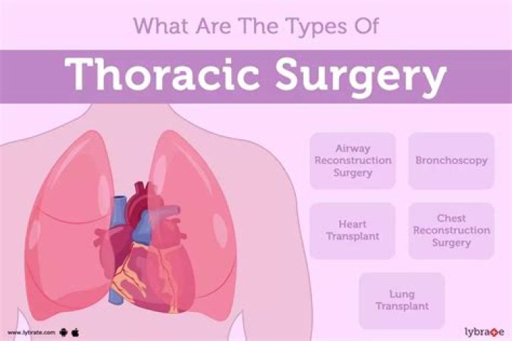
Who performs a thoracotomy? The following specialists perform a thoracotomy: Thoracic surgeons specialize in the surgical treatment of diseases of the chest, including the blood vessels, heart, lungs and esophagus. Thoracic surgeons may also be known as cardiothoracic surgeons.
What is a anterolateral thoracotomy?
The anterolateral thoracotomy provides excellent access to either upper lobe, the right middle lobe, and the anterior hila. It can be extended across the sternum into the opposite chest (clamshell incision). Anterolateral thoracotomy is our preferred approach for unilateral lung transplantation.
What's the meaning of pneumonectomy?
Listen to pronunciation. (NOO-moh-NEK-toh-mee) Surgery to remove all of one lung. In a partial pneumonectomy, one or more lobes of a lung are removed.
Can paramedics perform thoracotomy?
The 2003 guidelines for withholding or terminating resuscitation in prehospital traumatic cardiopulmonary arrest by NAEMSP and ACS said, “Thoracotomy is not a procedure that falls under the purview of prehospital care.”29 This may be true in a paramedic-run EMS system, as thoracotomy should not be a procedure expected …
Which muscles are cut in posterolateral thoracotomy?
In these procedures, it is common to perform a posterolateral thoracotomy with a division of the latissimus dorsi and serratus muscles, so it provides a good exposition and approach to any intrathoracic structure.
When was the first thoracotomy performed?
Historically the first successful Emergency Thoracotomy (ET) was carried out on September 14, 1902 on a kitchen table in Montgomery, Alabama.
Is a thoracotomy a chest tube?
Thoracostomy is a minimally invasive procedure in which a doctor inserts a thin plastic tube into the pleural space — the area between the chest wall and lungs. They may attach the tube to a suction device to remove excess fluid or air. Or, they may use the chest tube to deliver medications into the pleural space.
When is thoracotomy used in Hemothorax?
Thoracotomy is the procedure of choice for surgical exploration of the chest when massive hemothorax or persistent bleeding is present. At the time of surgical exploration, the source of bleeding is controlled and the hemothorax is evacuated.
When is a thoracotomy performed?
Thoracotomy is indicated when total chest tube output exceeds 1500 mL within 24 hours, regardless of injury mechanism. THE INDICATIONS for thoracotomy after traumatic injury typically include shock, arrest at presentation, diagnosis of specific injuries (such as blunt aortic injury), or ongoing thoracic hemorrhage.
What is superior facet?
The superior costal facet (or superior costal fovea) is a site where a rib forms a joint with the top of a vertebra. Ribs connect to the thoracic vertebrae at two main points, the inferior and superior costal facets. These connection points are located on two different vertebrae that are located on top of one another.
What is articular facet?
A facet is a flat or nearly flat surface on a bone. The vertebral articular facets are where two vertebrae articulate. There will be one pair of facets on the superior side of the vertebrae and one pair on the inferior side of the vertebrae.
What is sacral vertebra?
The sacral vertebrae—also called the sacral spine—consists of five sacral vertebrae bones. These bones fuse together to form the sacrum, the shield-shaped bony structure located at the base of the lumbar vertebrae (the five cylindrical bones forming the spine of the lower bank) and connected to the pelvis.
Why is L5 atypical?
The only atypical vertebra of the lumbar region is L5. L5 has the largest vertebral body and transverse processes. … The anterior aspect of the vertebral body is higher than the posterior aspect, contributing to the slightly wedge-shaped appearance it has.
What makes Atlas atypical?
C1 and C2 are considered atypical vertebrae because they have some distinguishing features compared to the rest of the cervical spine. C1 Vertebra (the atlas). The top vertebra, called the atlas, is the only cervical vertebra without a vertebral body. Instead, it is shaped more like a ring.
Why is C7 atypical?
C7 may be considered typical or atypical but has two distinct features. The first is that unlike the rest of the cervical vertebrae, is that the vertebral artery does not traverse the transverse foramen. The second is that it contains a long spinous process, also known as “vertebra prominens.”
What is foramen Transversarium?
The transverse foramen (foramen transversarium) of cervical vertebrae is an opening that is occupied by the vertebral artery and vein in the first six vertebrae and only the vertebral vein in the seventh. … These foramina do not occur in the other regions of the vertebral column.
What is superior to C1?
Superiorly, the C1 vertebra articulates (forms a joint) with the occipital condyles of the skull. … Below these are the 12 thoracic vertebrae, designated T1–T12. The lower back contains the L1–L5 lumbar vertebrae. The single sacrum, which is also part of the pelvis, is formed by the fusion of five sacral vertebrae.
What is C1 vertebra called?
Atlas (C1) The atlas is ring-shaped and does not have a body, unlike the rest of the vertebrae. Fused remnants of the atlas body have become part of C2, where they are called the odontoid process, or dens.
How long does it take to fully recover from a thoracotomy?
In 4-6 weeks you should be back to full activity and feel more like yourself. Take a couple of short walks outside each day (unless the weather is bad). Walking is excellent exercise. Taking deep breaths while walking will increase your strength.
Do people survive thoracotomy?
The primary outcomes analyzed were in-hospital survival rates. Results: EDT had an overall survival rate of 7.4%. Normal neurologic outcomes were noted in 92.4% of surviving patients. Factors reported as influencing outcomes were the mechanism of injury (MOI), location of major injury (LOMI), and signs of life (SOL).
How long does it take to recover from thoracic surgery?
Once home, patients who’ve undergone a minimally invasive surgery for lung cancer typically regain most of their strength, stamina, energy level, and breathing after two to three weeks. For open surgery, this level of recovery may take three to five weeks.