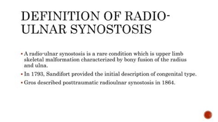
Radioulnar synostosis is a rare condition in which the two bones of the forearm — the radius and the ulna — are abnormally connected. This limits rotation of the arm.
Can radioulnar synostosis be fixed?
If your child has radioulnar synostosis in both arms, or if their forearm is fixed in a position that limits the function of their arm, they may benefit from surgery. Surgery is usually performed before children reach school age. Surgery involves repositioning the forearm so children can improve the use of the arm.
Is bilateral radioulnar synostosis a disability?
The condition can lead to significant disability, especially if there is hyperpronation or when it is bilateral, as occurs in 50% to 80% of cases.
How many cases of radioulnar synostosis are there?
Congenital. Congenital radioulnar synostosis is rare, with approximately 350 cases reported in journals, and it typically affects both sides (bilateral) and can be associated with other skeletal problems such as hip and knee abnormalities, finger abnormalities (syndactyly or clinodactyly), or Madelung’s deformity.Is Synostosis a disability?
If you or your dependent(s) are diagnosed with Congenital Radioulnar Synostosis and experience any of these symptoms, you may be eligible for disability benefits from the U.S. Social Security Administration.
What is Madelung deformity?
Madelung’s deformity is a rare arm condition that affects the growth plate of the radius, a bone in the forearm. As a child grows, this abnormal growth results in a misalignment where the two long bones of the forearm (the radius and ulna) meet the bones of the wrist.
What is the radioulnar syndesmosis?
Description. Radioulnar syndesmosis is a slightly movable articulation of the forearm where the contiguous bony surfaces from radius and ulna are united by interosseous ligaments : the interrosseous membrane of forearm and the oblique cord.
What are the symptoms of craniosynostosis?
- A full or bulging fontanelle (soft spot located on the top of the head)
- Sleepiness (or less alert than usual)
- Very noticeable scalp veins.
- Increased irritability.
- High-pitched cry.
- Poor feeding.
- Projectile vomiting.
- Increasing head circumference.
Does Radioulnar have Synostosis?
Congenital radioulnar synostosis is a rare condition in which the forearm bones (radius and ulna) are fused together at the elbow, preventing a child from rotating their palm up or down. The condition is often present in both arms. Left untreated, it can result in limited function.
What causes congenital radial head dislocation?The etiology of congenital radial head dislocations is believed to be an abnormal embryologic development of the capitulum. This abnormality results in the loss of contact with the radial head, which subsequently develops into a malformation of the radiocapitellar joint, the radioulnar joint, and the ulnar head.
Article first time published onWhat's a synostosis?
Medical Definition of synostosis : union of two or more separate bones to form a single bone also : the union so formed (as at an epiphyseal line) Other Words from synostosis. synostotic \ -ˈtät-ik \ adjective.
Is radioulnar joint syndesmosis?
The interosseous membrane of the forearm (rarely middle or intermediate radioulnar joint) is a fibrous sheet that connects the interosseous margins of the radius and the ulna. It is the main part of the radio-ulnar syndesmosis, a fibrous joint between the two bones.
What is congenital proximal radioulnar synostosis?
Congenital proximal radioulnar synostosis is a rare malformation of bone development characterized by the fusion of the proximal radius and ulna. This malformation usually occurs bilaterally and is diagnosed before the patient is 5 years old.
Where is the distal radioulnar joint?
Distal radioulnar joint – located near the wrist. It is an articulation between the ulnar notch of the radius and the ulnar head.
What is ulna and radius?
The radius and the ulna constitute as the bones of the forearm. The antebrachial region, as it is clinically known, spans the length of the region which extends roughly from elbow to wrist. The radius is the lateral of the two bones, which makes the ulna the medial bone of the forearm.
What causes sagittal synostosis?
Sagittal craniosynostosis occurs when certain bones in a child’s skull fuse prematurely. At birth, a child’s skull is made up of several separate bones with growth plates between them. Because the skull is not a solid piece of bone yet, the brain can grow and expand in size.
What is sagittal synostosis?
Sagittal synostosis (scaphocephaly) is the premature closure of the sagittal suture of the skull that causes abnormal growth of the skull resulting in a long and narrow head shape with fullness (bossing) of the forehead.
What causes Scaphocephaly?
Scaphocephaly is caused by the early fusion of the sagittal suture which runs from front to back at the top of the skull. Early fusion of a suture in infancy is called synostosis and this type is the most common form of craniosynostosis.
What is superior Radioulnar?
Anatomical terminology. The proximal radioulnar joint (superior radioulnar joint) is a synovial pivot joint between the circumference of the head of the radius and the ring formed by the radial notch of the ulna and the annular ligament.
What is the pronator quadratus?
Pronator quadratus is a quadrangular, thin, short and flat muscle lying within the anterior compartment of forearm. It is part of the deep group of forearm flexors, together with flexor digitorum profundus and flexor pollicis longus. … Pronator quadratus extends across the distal parts of the radius and ulna.
What fits in the Coronoid fossa?
Anatomical terms of bone Superior to the anterior portion of the trochlea is a small depression, the coronoid fossa, which receives the coronoid process of the ulna during flexion of the forearm. It is directly adjacent to the radial fossa of the humerus.
What is the bone in your wrist that sticks out called?
Pisiform boneMeSHD051220TA98A02.4.08.007TA21254FMA23718
What is a dinner fork deformity?
A dinner fork deformity, also known as a bayonet deformity, occurs as the result of a malunited distal radial fracture, usually a Colles fracture. The distal fragment is dorsally angulated, displaced and often also impacted.
What is made lung?
Madelung deformity refers to bowing of the radial shaft with increased interosseous space and dorsal subluxation of the distal radioulnar joint. This deformity is due to premature closure or defective development of the ulnar third of the distal physis of the radius.
How do you test for craniosynostosis?
Doctors can identify craniosynostosis during a physical exam. A doctor will feel the baby’s head for hard edges along the sutures and unusual soft spots. The doctor also will look for any problems with the shape of the baby’s face.
Where is synostosis located?
Synostoses may occur between all or any two of the three bones present at the elbow. The most common synostosis is that between the radius and the ulna proximally in the forearm, near the elbow (Fig.
What is Lambdoid synostosis?
What Is Lambdoid Synostosis? This condition occurs when the bones at the back of an infant’s skull close up or fuse together prematurely. Normally, the bones of the skull close after reaching adulthood. With lambdoid synostosis, the bones at the base of the skull fuse too soon.
What is adult craniosynostosis?
Introduction. Craniosynostosis refers to closure of calvarial sutures prematurely resulting in restricted skull growth. It is classified as primary and secondary. The patient presents with unexplained neuropsychological impairment. Radiological imaging is necessary for establishing the diagnosis.
What age does craniosynostosis occur?
When the bones of the skull are fused together either at birth or fuse too soon, the condition is called craniosynostosis. The sutures of the skull fuse around the brain at around age 2 years. When a baby has craniosynostosis, one or more of these sutures hardens too early and closes before the baby reaches age 2.
What is radial head dislocation?
Radial head dislocation occurs when the radial head is displaced from its normal articulation with the ulna and the humerus. The dislocation may be acquired or congenital (see the separate article on congenital radial head dislocation).
What is ulnar dysplasia?
Babies with ulnar dysplasia (also called ulnar club hand) are born with a short or missing ulnar bone. The ulnar bone is one of the two forearm bones. The short or missing ulnar bone causes the hand and wrist to turn outward toward the pinky side of the forearm. The hand, wrist, and elbow can have changes too.