RodsConesConfer achromatic visionConfer color vision
What is rod cells and cone cells?
There are two types of photoreceptors in the human retina, rods and cones. Rods are responsible for vision at low light levels (scotopic vision). … Cones are active at higher light levels (photopic vision), are capable of color vision and are responsible for high spatial acuity.
Which are called as the nerve cells of retina?
Anatomical terms of neuroanatomy A retinal ganglion cell (RGC) is a type of neuron located near the inner surface (the ganglion cell layer) of the retina of the eye. It receives visual information from photoreceptors via two intermediate neuron types: bipolar cells and retina amacrine cells.
What type of neurons are rods and cones?
1. Photoreceptors There are two main types of light-sensitive cell in the eye: rods and cones. Rods enable vision in poor light, whereas cones are responsible for colour vision. Photoreceptors convert light into electrical signals that travel through other retinal neurons to reach the optic nerve.Are rods and cones ganglion cells?
In most parts of the retina, rod and cone signals converge on the same ganglion cells; i.e., individual ganglion cells respond to both rod and cone inputs, depending on the level of illumination. The early stages of the pathways that link rods and cones to ganglion cells, however, are largely independent.
What is the chemical and difference between rods and cones?
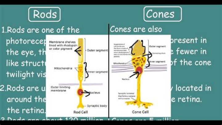
RodsCones2They have visual purple pigment called as rhodopsin.They have visual violet pigment called as iodopsin.3Rods are the photoreceptor cells of the retina that are sensitive to dim light.Cones are the photoreceptor cells of the retina that are sensitive to bright light.
How are rod cells and cone cells different?
Photoreceptor cells Photoreceptors in the retina are classified into two groups, named after their physical morphologies. Rod cells are highly sensitive to light and function in nightvision, whereas cone cells are capable of detecting a wide spectrum of light photons and are responsible for colour vision.
Are cones cells?
Cone cells, or cones, are photoreceptor cells in the retinas of vertebrate eyes including the human eye. … Cones are less sensitive to light than the rod cells in the retina (which support vision at low light levels), but allow the perception of color.Are rods and cones epithelial cells?
RodsConesConfer achromatic visionConfer color vision
What is the nerve cell?(nerv sel) A type of cell that receives and sends messages from the body to the brain and back to the body. The messages are sent by a weak electrical current. Also called neuron.
Article first time published onWhat are glial cells?
Glial cells, also called glial cells or neuroglia, are cell which are non-neuronal and are located within the central nervous system and the peripheral nervous system that provides physical and metabolic support to neurons, including neuronal insulation and communication, and nutrient and waste transport.
Are ganglion cells neurons?
Overview. Ganglion cells are the final output neurons of the vertebrate retina. Ganglion cells collect information about the visual world from bipolar cells and amacrine cells (retinal interneurons). This information is in the form of chemical messages sensed by receptors on the ganglion cell membrane.
Why are rods and cones at the back of the retina?
On the retina, the back of the eye, the light rays pass right through the nerve cells that will pass signals to the brain—but ignore them for now. They reach cones—that line the back of the eye and sense the differences in colors—and rods, which are color-blind but even more sensitive to light.
Where are rod and cone cells located?
They are located in the retina (a layer at the back of the eye). There are two types, rods and cones.
What cells do rods and cones connect to?
Rods and cones make synapses in the outer plexiform layer with two kinds of cells ( Figure 1): bipolar cells, whose axons transmit information to the next layer of retina called the inner plexiform layer, and horizontal cells, whose lateral processes interconnect photoreceptors and bipolar cells.
What are the functions of rods and cones?
Rods and cones are the receptors in the retina responsible for your sense of sight. They are the part of the eye responsible for converting the light that enters your eye into electrical signals that can be decoded by the vision-processing center of the brain.
What is the difference between rods and cones quizlet?
Rods are ultra-sensitive to light and simply detect light, good for night vision. … Cones are responsible for color vision.
What is the function of rod cells quizlet?
Rod cells, or rods, are photoreceptor cells in the retina of the eye that can function in less intense light than the other type of visual photoreceptor, cone cells. Rods are concentrated at the outer edges of the retina and are used in peripheral vision.
How do rod cells work?
Rod cells function as specialized neurons that convert visual stimuli in the form of photons (particles of light) into chemical and electrical stimuli that can be processed by the central nervous system. … Rod cells are much more sensitive to light than cones and are also much more numerous.
How cones and rods are distributed in retina?
Distribution of rods and cones in the human retina. Graph illustrates that cones are present at a low density throughout the retina, with a sharp peak in the center of the fovea. Conversely, rods are present at high density throughout most of the retina, (more…)
Is Cone a nerve cell?
cone, light-sensitive cell (photoreceptor) with a conical projection in the retina of the vertebrate eye, associated with colour vision and perception of fine detail. … Chemical changes that occur when light strikes the cones are ultimately relayed as impulses to optic-nerve fibres that enter the brain.
What type of receptor is a rod cell?
Rod cellFunctionLow-light photoreceptorNeurotransmitterGlutamatePresynaptic connectionsNonePostsynaptic connectionsBipolar cells and horizontal cells
Are the cones in the macula?
The Center of Vision: The Macula The retina, located in the back of the eye, contains several layers of photoreceptor cells (known as rods and cones).
What are 3 types of nerve cells?
- Sensory neurons. …
- Motor neurons. …
- Interneurons. …
- Neurons in the brain.
What are the 4 types of cells?
- Epithelial Cells. These cells are tightly attached to one another. …
- Nerve Cells. These cells are specialized for communication. …
- Muscle Cells. These cells are specialized for contraction. …
- Connective Tissue Cells.
Are neuron cells?
A neuron or nerve cell is an electrically excitable cell that communicates with other cells via specialized connections called synapses. It is the main component of nervous tissue in all animals except sponges and placozoa. … A typical neuron consists of a cell body (soma), dendrites, and a single axon.
Are glial cells nerves?
Glial cells are smaller than neurons but are greater in number than nerve cells in the brain. Glial cells do not have axon and dendrites. However, they come into play during neural development or recovery from neural injury and during modulation of synaptic action and propagation of nerve signals.
What are the two types of glia cells?
There are three types of glial cells in the mature central nervous system: astrocytes, oligodendrocytes, and microglial cells (Figure 1.4A—C). Astrocytes, which are restricted to the brain and spinal cord, have elaborate local processes that give these cells a starlike appearance (hence the prefix “astro”).
What are the 6 glial cells?
Neuroglia in the CNS include astrocytes, microglial cells, ependymal cells and oligodendrocytes. In the PNS, satellite cells and Schwann cells are the two kinds of neuroglia.
What is ganglion nerve?
A ganglion is a collection of neuronal bodies found in the voluntary and autonomic branches of the peripheral nervous system (PNS). Ganglia can be thought of as synaptic relay stations between neurons. The information enters the ganglia, excites the neuron in the ganglia and then exits.
Are ganglion cells bipolar?
Ganglion cells receive synaptic input from as few as one or as many as a hundred bipolar cells depending on their eccentricity. Their axons project across the inner surface toward the center of the retina, converging at the optic disc.